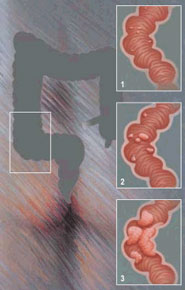
What Is Colon Cancer?
The phrase “colon cancer” includes various epithelial tumours depending on the histologic structure, shape, location (cecum, ascending colon, transverse colon, sigmoid colon, rectum and anal canal).
Colon Cancer Etiology
It seems that there is no single cause for colon cancer. It is most probably caused by a combination of harmful (unfavourable) factors, of which the most important ones are the diet, the environmental factors, the chronic colon diseases and the heredity.
Several scientists and medical trials focus on role of the diet in the colorectal cancer pathogenesis. Colorectal cancer is more frequently encountered in areas in meat-predominating diets and low vegetable consumption. A meat-rich diet leads to the increase of the concentration of fatty acids that are transformed into carcinogens during the digestion process. The low colorectal cancer rate in traditionally vegetable-based diet areas (India, Central Africa) demonstrates the importance of a diet rich in vegetal fibres in the prevention of colon cancer. The low vegetal fibre intake increases the fecal mass volume, dilutes and binds various carcinogens, shortens the colon transition time, thus reducing the colon mucous membrane exposure time to the action of carcinogenic metabolites.
The chemical theory is similar to the nutritional theory. It explains the onset of colorectal tumours as a result of the mutagenic action of certain endo- and exogenous (carcinogenic) factors on the colon epithelium cells, of which the most dangerous and active ones are the nitrates, nitrites, amides and aromatic amines, alpha toxins, tryptophan and tyrosine metabolites. Carcinogenic substances (benzopyrene) are obtained through the irrational thermal processing of food, such as meat and fish smoking. As a result of the action of the carcinogenic substances on the cell genome, mutations, translocations occur, leading to the transformation of cell proto-oncogenes into active oncogenes. The latter initiate the synthesis of the oncoproteins and the cell transforms into a cancer cell.
In patients with inflammatory colon diseases, especially with ulcero-hemorrhagic rectocolitis, the colon cancer incidence is much higher than in the rest of the population. The risk degree is influenced by the duration and clinical evolution of the disease, the older the condition, the higher the cancer risk. In Crohn’s disease, the colon cancer risk is still high, but lower than in RCUH.
Colon polyps increase the malign tumour risk. The solitary polyp subsequent malignancy risk is of 2-4%, whereas for multiple polyps (more than 2) it is of 20%, and in villous polyps it is as high as 40%. Colon polyps are relatively rare in the young population and much more frequent in the elderly. The frequency of colon polyps may be established by analyzing the autopsy data. In developed countries, the colon polyp identification frequency upon autopsies performed in persons deceased for non-colon-related causes is of 30-32%.
Heredity also plays an important role in the pathogenesis of colon cancer. People who have first degree relatives suffering from malign colon tumours are exposed to a high risk. Hereditary diseases, such as diffuse familial adenomatous polyposis, Gardner’s syndrome, Turcot’s syndrome have a high colon cancer risk. If the polyps or colon of these patients are not removed, almost all of them develop colon cancer, some with synchronous tumours.
The familial cancer syndrome, with autosomal dominant transmission, manifests through the appearance of multiple colon tumours (adenocarcinoma). Almost 1/3 of these patients aged above 50 feature colorectal tumours.
From amongst the peculiarities of the colon cancer, its multicentric development should be mentioned, with tumours developing at the same time (synchronous tumours) or one after another at certain time intervals (metachronous tumours).
Colon Cancer Clinical Presentation:
The most specific colorectal cancer symptoms are rectal bleeding, intestinal transit disorders, abdominal pain and tenesmus.
Rectorrhagia, faeces mixed with blood or the identification of occult bleeding are practically present in all colorectal cancer patients. Bright red bleeding is specific to anal canal and rectum tumours. Dark coloured blood suggests a tumour on the left hand-side of the colon. It should be mentioned that the blood mixed with faeces and mucus is an almost certain sign of tumour. Right-hand side tumours are characterized by occult bleeding, associated to anaemia, pale teguments and general weakness.
Transit disorders are specific to the advanced forms of right-hand side colon cancer. Colon cancer sometimes directly manifests through intestinal obstruction, which requires an emergency medical treatment.
In rectum cancer, patients accuse a feeling of incomplete defecation or a false defecation sensation. Abdominal symptoms are often absent, the sensation of weakness, the lack of appetite, the weight loss being more frequently encountered. In tardy stages, it is associated to hepatomegaly and ascites.
How Is Colon Cancer Diagnosed?
The current medical solutions allow for the diagnosis of colon cancer in all stages of the disease. Two conditions must be observed:
- the investigation algorithm;
- the maximum use of all available modern investigation means
Colon cancer diagnosis algorithm includes:
- the analysis of the symptoms and the anamnesis (remember that the colon cancer risk significantly increases in people aged above 50);
- clinical examination;
- rectal examination;
- anoscopy;
- rectocolonoscopy;
- blood tests;
- analysis of faeces for occult blood;
- colonoscopy;
- biopsy of identified tumours or polyps;
- irrigoscopy (if colonoscopy is not conclusive or is missing);
- abdominal and pelvic ultrasound;
- transrectal ultrasound;
Colon disease-specific symptoms are somehow monotonous. Most involve transit disorders, the presence of blood and mucus in the faeces or abdominal and rectal pain or discomfort. These symptoms are often combined.
There is no specific colorectal cancer sign. Hence, all intestinal symptoms must be analyzed as a potential manifestation of colorectal cancer, especially in people aged above 50. Most tumours (up to 70%) are located in the distal area of the colon (rectum and sigmoid). For instance, in order to identify a distal rectal ampulla tumour, a simple rectal examination is sufficient. In order to take maximum advantage of all diagnosis possibilities, the adequate preparation of the colon is required. If not, diagnosis errors by omission are possible.
An important diagnosis method is ultrasound, which can show not only the presence of distal (especially hepatic) metastases, but also the local invasion degree of the tumour and the presence of the perifocal inflammation. In advanced cases, with invasion in the neighbouring organs and tissues, computed tomography or nuclear magnetic resonance imaging is recommended.
Colon Cancer Complications:
The most often encountered colon cancer complications are intestinal transit disorders up to intestinal obstruction, intestinal bleeding, perifocal inflammations and intestine or tumour-level perforations, or the so-called dilation perforations in the case of intestinal occlusions. In the case of right-hand side colon tumours, severe anaemia is also frequent, because of the long-term occult bleeding.
All complication require suitable treatment, sometimes even surgical interventions to save the patient’s life, such as it is the case with important bleeding, obstructions or perforations. In the advanced colorectal cancer cases, complications may combine, increasing the risks and aggravating the surgical intervention prognosis. The solution to prevent complications is the early diagnosis of colorectal cancer.
How Is Colon Cancer Treated?
Surgical intervention continues to be the main method in the treatment of colorectal cancer. The general surgery principles are the radical nature of the surgical intervention, the ablation, the asepsis and the maintenance of the free intestinal transit, if naturally possible.
The surgical intervention is associated to an adjuvant and neoadjuvant treatment. Pre-surgery radiotherapy has a cytoreductive effect, i.e., it is meant to reduce the volume of the tumour and ensure the maximum radicality of the surgery in combination with the locoregional lymphadenectomy. Post-operative chemotherapy is meant to destroy the cancer cells and prevent local relapse or metastases.
The presence of distal metastases, in the liver or other organs, imposes, in addition to the ablation of the tumour and the resection for cytoreductive, the post-surgical chemotherapy. Radio and chemotherapy schemes are well defined depending on the type of tumour identified.
Colon Cancer Prognosis:
Colon cancer prognosis depends upon the stage of the disease. In the incipient stages (T, N0, M0) the survival for 5 years post-intervention may be as high as 90% . However, in more advanced stages of the disease, the results decrease. In patients with ganglion implications, the 5-year survival rate does not exceed 50%, while in the cases right-hand side colon location it does not exceed 20%. The long-term survival results of patients suffering from rectal cancer are poorer. As an average, 5-year survival in patients with radical rectal cancer surgeries is of 50% and it also depends on the stage of the process.
Regular follow-up visits are mandatory in patients with colorectal cancer surgeries, so as to allow for the early identification of the local relapses or possible metastases. A rectal examination is recommended every 3 months, as well as the colonoscopy or irigography remaining colon segment after the resection, abdominal, hepatic and pelvic ultrasound every 6 months, pulmonary RX. The titration of the carcinoembryonic antigen from the tests is recommended. In the case of suspected relapses, the computed tomography or magnetic resonance imaging is required.
85% of the local relapses occur during the first 2 years after the surgery, but the average term is of 13 months. A new resection is possible in approximately 1/3 of patients with early identified relapse. For the rest, we must regretfully admit that palliative care (radio- and chemotherapy) that slightly improves the patients’ condition is the only solution.


