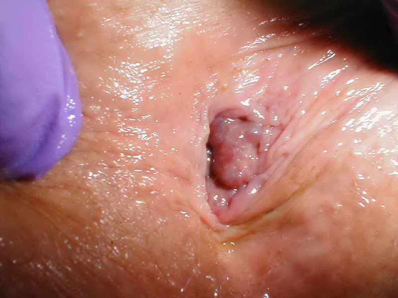
 Colon Polyps are clearly delimited growths (protrusions), developed at the level of the colon mucosa. These lesions are very important, because they are regarded as pre-cancerous growths, responsible for most colorectal cancer forms. The identification and endoscopic resection of these lesions, as well as the patients’ follow-up, decrease or even remove the individual colorectal cancer risk. Colon polyps vary in shape, size and location.
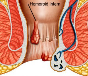
They can be sessile, with a wide implantation basis or pedunculated, with a pedicle (foot) that links the polyp head to the colon wall. Depending on their size, they can be small (below 5mm), average (5-15mm) and large (above 15mm), the latter featuring a high malignant transformation risk. Polyps can occur anywhere at the level of the colon, but there is, however, a predisposition for the left (descendent, sigmoid) colon in 70% of the cases. Hence, flexible rectosigmoidoscopy is an excellent and cost-efficient method to detect these lesions.
The histologic substrate of colon polyps is varied, determining the differences with regards to the natural history, evolution and prognosis. In brief, polyps can be “adenomatous” (tubular, villous or tubulovillous), characterized through variable glandular atypia and a potential of evolution towards colorectal cancer, and “non-adenomatous” (hyperplasic), with minor clinical consequences. Hence, the correct approach of these lesions always starts from the anatomic-pathological (microscopic) examination.
The transformation of adenomatous polyps into cancer is an unpredictable process, with a variable duration (between 2 and 10 years), the risk increasing with the size, the histological type, the number of polyps, the patient’s age. Malignization is only one of the possible evolutions of a polyp and it is characterized by the accumulation of successive genetic mutations in the glandular epithelium cells.
The incidence of colon polyps in endoscopic studies increases with the age, from 10% at 45 to 40-50% in people aged above 55-60. Of those, more than 75% are adenoma, especially for those with sizes above 5mm.
Methods of diagnosis:
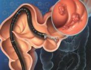
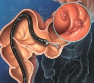
Most often, colon polyps are diagnosed by chance as part of the colonoscopy examinations, as the patients are asymptomatic. Sometimes, large polyps, above 10mm, distally located, can cause bleeding (rectorrhagia), which must be regarded as a warning sign. That is why colonoscopy is mandatory in the case of any rectal bleeding, in order to set a diagnosis. Unlike other methods, colonoscopy allows for a detailed description of the polyps, offering details on its location, size, shape, aspect of the mucous membrane and also helps prevent microscopic examination biopsy collection. Intestinal transit changes (diarrhea and/or constipation) can occur in the case of large polyps, distally located.
Methods of treatment:
Endoscopic polypectomy (polyp excision during colonoscopy) represents the optimum method for the treatment of colorectal polyps. Polypoid lesions must be fully excised and recovered for the histology examination. Moreover, the endoscopic examination of the entire colon is required in order to identify the possible associated lesions. Sometimes, the follow-up colonoscopies are required 1 year after the polypectomy and at individualized intervals thereafter depending on the peculiarities of the excised polyps.
Laurus Medical Clinics throughout the country, offer state-of-the-art equipment rectosigmoidoscopy, performed by gastroenterologists with experience in diagnosis and therapeutic GI endoscopy. An internal review of a number of 7,000 patients investigated and treated in our clinic has shown that flexible rectosigmoidoscopy is a safe and efficient diagnosis procedure, causing minimum discomfort to the patient. A number of 740 patients were diagnosed with colon polyps (10.5%), of whom 190 had multiple polyps. An overall number of 980 polyps were identified and biopsied, and an important percentage was endoscopically removed. Moreover, 80 patients (1.15%) were diagnosed with colorectal cancer and subsequently treated in university clinics. No incidents were registered during the rectosigmoidoscopy.
To conclude, colonoscopy continues to be the ideal method to identify and remove colorectal polyps, as it offers an excellent sensitivity and specificity, and thus considerably reduces the colorectal cancer risk.

